KAIST
BREAKTHROUGHS
Research Webzine of the KAIST College of Engineering since 2014
Spring 2026 Vol. 26Simultaneous magnetic resonance arteriography and venography
A new method for simultaneous acquisition of a magnetic resonance (MR) arteriogram and an MR venogram is developed. The approach provides clinicians with diverse vascular information within a short scan time, which is crucial for clinical diagnosis of patients, especially under time-sensitive conditions such as stroke.
Article | Fall 2019
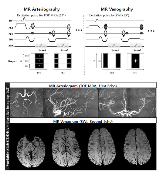
Magnetic resonance (MR) arteriogram and MR venogram are routinely acquired in hospitals for clinical diagnosis, typically using time-of-flight MR angiography (TOF MRA) and susceptibility-weighted imaging (SWI), respectively. Despite the necessity of both, only one of them more relevant to the clinical diagnosis is selectively acquired in practice because of their long scan times. There have been trials to acquire MR arteriogram and venogram simultaneously. However, none of them has been widely used either because the scan conditions for one of the two scans are far from the optimal condition or because the scan time gets longer; therefore, the advantage of the simultaneous acquisition becomes meaningless.
A research team of Associate Professor Sung-Hong Park from the Department of Bio and Brain Engineering at the Korea Advanced Institute of Science and Technology (KAIST) addressed this issue. The research team focused on the fact that MR image acquisition is performed on the frequency domain, where the low frequency component stores most of the energy and determines the anatomical contrast of MR images. They developed a special dual-echo imaging technique termed “variable-slab compatible dual-echo arteriovenography” (vCODEA ), to acquire MR arteriogram (first echo) and venogram (second echo) simultaneously. In contrast to the previous techniques, the team manipulated the acquisition order of the two echoes so that the low frequency components of the first and second echoes were acquired at different magnetic resonance shots (radio frequency excitations) with no change in the total scan time. The optimal scan conditions for the MR arteriography (multiple overlapping thin‑slab acquisition, ramped‑profile excitations, higher flip angle, etc.) were then applied during the acquisition of the low frequency component of the first echo, whereas those for the MR venography (single thick‑slab acquisition, flat-profile excitations, lower flip angle, etc.) were applied during the acquisition of the low frequency component of the second echo. The team also demonstrated that the proposed technique can be combined with a parallel imaging technique to decrease scan time further.
A noteworthy outcome of this study was that we can now truly acquire optimal MR arteriogram and optimal MR venogram simultaneously in a scan time corresponding to only one of the two scans. The proposed approach has additional benefit of decreasing slab-boundary artifacts for the MR arteriography, which have been problematic in typical multi-slab acquisitions.
The proposed MR arteriovenography will be useful for clinical diagnosis of patients, especially under time-sensitive conditions such as stroke. The technique can provide clinicians with diverse vascular information with optimal scan conditions within a limited scan time.
This research was published in IEEE Transactions on Medical Imaging (CiteScore Rank: Top 2%~4% depending on categories) (Do WJ, Choi SH, Park SH. IEEE-TMI; 2018;37(7):1632-1640. DOI: 10.1109/TMI.2018.2789923).
Most Popular

Lighting the Lunar Night: KAIST Develops First Electrostatic Power Generator for the Moon
Read more
Soft Airless Wheel for A Lunar Exploration Rover Inspired by Origami and Da Vinci Bridge Principles
Read more
GPU-NPU-PIM Integration Technology for Generative AI Clouds
Read more
Dual-Action Hydrogel Offers New Hope for Rheumatoid Arthritis Treatment
Read more
Title WSF1 Vision Concept: Redefining Wearable Robotics through Human-centred Mobility Design
Read more